


University of Kansas
School of Medicine
Kansas City
In this month’s column, I highlight three topics that are highly relevant to endoscopy clinical practice: colon polyp sizing, Helicobacter pylori’s relationship with gastric cancer and large nonpedunculated polyps.
Although pathologists report the size of colon polyps resected during colonoscopy, the accuracy of such size reporting is questionable. In this study, investigators showed that due to factors such as piecemeal resection, only 60% of polyps sent to pathology can be measured, and of those that are measured, shrinkage of polyp size occurs in almost 25% of the specimens.
The majority of the results showing a reduction in gastric incidents after H. pylori incidence—a known risk factor for noncardia gastric adenocarcinoma (NCGA)—are from data collected outside the United States. In the second study, a large population-based study from the United States, investigators show a continued significant risk reduction eight years later in NCGA after H. pylori eradication.
Large nonpedunculated colonic polyps (LNPCPs) seen during colonoscopy are associated with synchronous lesions in up to 60% of patients. In the third study I highlight, investigators report the characteristics and morphology of such lesions and show that a higher rate of synchronous lesions is found in large nongranular nonpedunculated colonic polyps.
Polyp Measurements in Pathology
Gut 2023 July 28. doi:10.1136/gutjnl-2023-330241
In this prospective clinical study, researchers in Canada evaluated the accuracy of colorectal polyp size measurements during pathology.
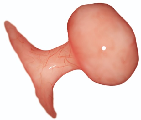
The researchers reviewed 482 resected polyps in 203 consecutive patients undergoing screening or diagnostic colonoscopy. Only 286 (59%) of the resected polyps were evaluated for size due to piecemeal resection or fragmentation during the retrieval process.
Polyps originally were measured for size during colonoscopy using a laser-based virtual scale endoscope (VSE) (EW10-VM01 for Scale Eye, Fujifilm). After removal, viable specimens were measured before formalin fixation using a digital Vernier caliper (eSynic with 32 feeler gauge measuring with 0.01-mm intervals) and a magnification lamp (Veemagni 8’ LED lighted) to establish a reference size measurement.
In the measurements made during pathology, 78% were smaller than the original polyp size, indicating formalin fixation induces significant shrinkage of colorectal polyps. The relative accuracy of the intracolonoscopy VSE size measurements was significantly higher than the relative accuracy of pathology sizing (84.2% vs. 74.1%; P<0.0001).
The investigators concluded that the usefulness for pathology in determining polyp size is limited because about 40% of colorectal polyps cannot be removed en bloc, retrieved intact or processed adequately.
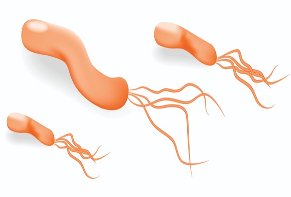
H. pylori Eradication
Gastroenterology 2023;165(2):391-401
In a retrospective cohort study, researchers at Kaiser Permanente Northern California evaluated the incidence of NCGA after H. pylori eradication.
The study evaluated 716,567 individuals with a history of H. pylori testing and/or treatment between 1997 and 2015 and followed through Dec. 31, 2018.
The researchers found patients who were treated for H. pylori infection had a lower risk for NCGA than those who had remained untreated (treated: adjusted subdistribution hazard ratio [HR], 2.68; 95% CI, 1.86-3.86; untreated: HR, 6.07; 95% CI, 4.20-8.76).
Compared with those seen in the general population of Kaiser Permanente Northern California, standardized incidence ratios of NCGA steadily decreased after H. pylori treatment: 2.00 (95% CI, 1.79-2.24) at one or more years, 1.01 (95% CI, 0.85-1.19) at four or more years, 0.68 (95% CI, 0.54-0.85) at seven or more years, and 0.51 (95% CI, 0.38-0.68) at 10 or more years.
The researchers concluded that H. pylori eradication therapy was associated with a significantly reduced incidence of NCGA after eight years compared with no treatment. After seven to 10 years of follow-up, the risk in treated individuals became lower than that of the general population.
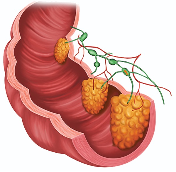
Synchronous Lesions
Clin Gastroenterol Hepatol 2023;21(9):2270-2277.e1
In this study, an international group of researchers examined the relationship between the surface morphology of LNPCPs and synchronous large lesions.
The investigators analyzed the Australian Colonic Endoscopic Resection cohort, a multicenter, observational study of consecutive patients referred for LNPCP resection. Patients were enrolled at one of seven sites with established resection programs over nearly 11 years until March 2022. The primary outcome of the study was the identification of individual lesion characteristics associated with the presence of synchronous LNPCPs. Serrated lesions and mixed granularity LNPCPs were excluded from the analysis, and LNPCP morphology was described according to the Paris classification.
In the study, 93.1% of patients had a single LNPCP. Those with solitary lesions had a median size of 35 mm, and lesions were predominantly located in the right colon (59.5%) and had a Paris 0 to IIa morphology (42.9%).
In the 6.9% of patients with two or more LNPCPs, the dominant lesion had a median size of 40 mm. Nearly half (47.6%) had a Paris 0 to IIa morphology, and the majority (65.9%) were in the right colon.
The researchers found that more dominant LNPCPs (35.8%) were nongranular (NG-LNPCP) in this group than in the solitary LNPCP cohort (18.7%). They also found that NG-LNPCPs were more likely to demonstrate synchronous disease, with left colon NG-LNPCPs (odds ratio [OR], 4.78; 95% CI, 2.95-7.73) demonstrating greater risk than right colon NG-LNPCPs (OR, 1.99; 95% CI, 1.39-2.86). Overall, NG-LNPCPs demonstrated a greater than fourfold increased risk for synchronous disease. Granularity, size and location influenced its likelihood.


This article is from the October 2023 print issue.