Virginia Commonwealth University
Richmond, Virginia
Stravitz-Sanyal Institute for Liver Disease and Metabolic Health
Virginia Commonwealth University
Richmond, Virginia
To address the diagnostic challenges of fibrosis staging in chronic liver disease (CLD), blood-based biomarkers and imaging-based modalities, collectively termed noninvasive liver disease assessments (NILDAs), have been developed to evaluate for the presence and degree of hepatic fibrosis, steatosis, and clinically significant portal hypertension.
Hepatic fibrosis is a significant disease threshold in chronic liver disease because most clinically relevant liver-related morbidity and mortality occurs in patients with advanced fibrosis (AF) or cirrhosis.1 In patients with fibrosis, hepatic decompensation and complications from portal hypertension drive poor outcomes. Fibrosis assessment is associated with prognosis and used to direct treatment strategies for hepatitis B virus (HBV), hepatitis C virus (HCV), and metabolic dysfunction–associated steatotic liver disease (MASLD). Therefore, fibrosis assessment to identify patients with moderate to AF and cirrhosis is crucial in the management of CLD.
Fibrosis stage classification ranges from F0 to F4, with fibrosis percentage increasing from F0 (normal) to F4 (cirrhosis).2 Degrees of fibrosis are stratified as at least significant fibrosis (=F2), at least AF (=F3), and cirrhosis (=F4).2-4 However, fibrosis staging is challenging because it is a nonlinear, dynamic process that is neither static nor necessarily progressive.
Traditionally, the diagnosis of CLD and fibrosis staging has relied on histologic evaluation by liver biopsy, but this approach has limitations. Liver biopsy is not only expensive and invasive but also is associated with adverse events, such as pain, bleeding, perforation of hollow viscus, and, rarely, death.5 Although liver biopsy is considered the gold standard in diagnosis of CLD, it can lead to incorrect fibrosis staging in up to 25% of cases due to length, number, and type of biopsies performed and etiology of liver disease.5
To address the diagnostic challenges in CLD, NILDAs have been developed to evaluate for the presence and degree of hepatic fibrosis, steatosis, and clinically significant portal hypertension.2
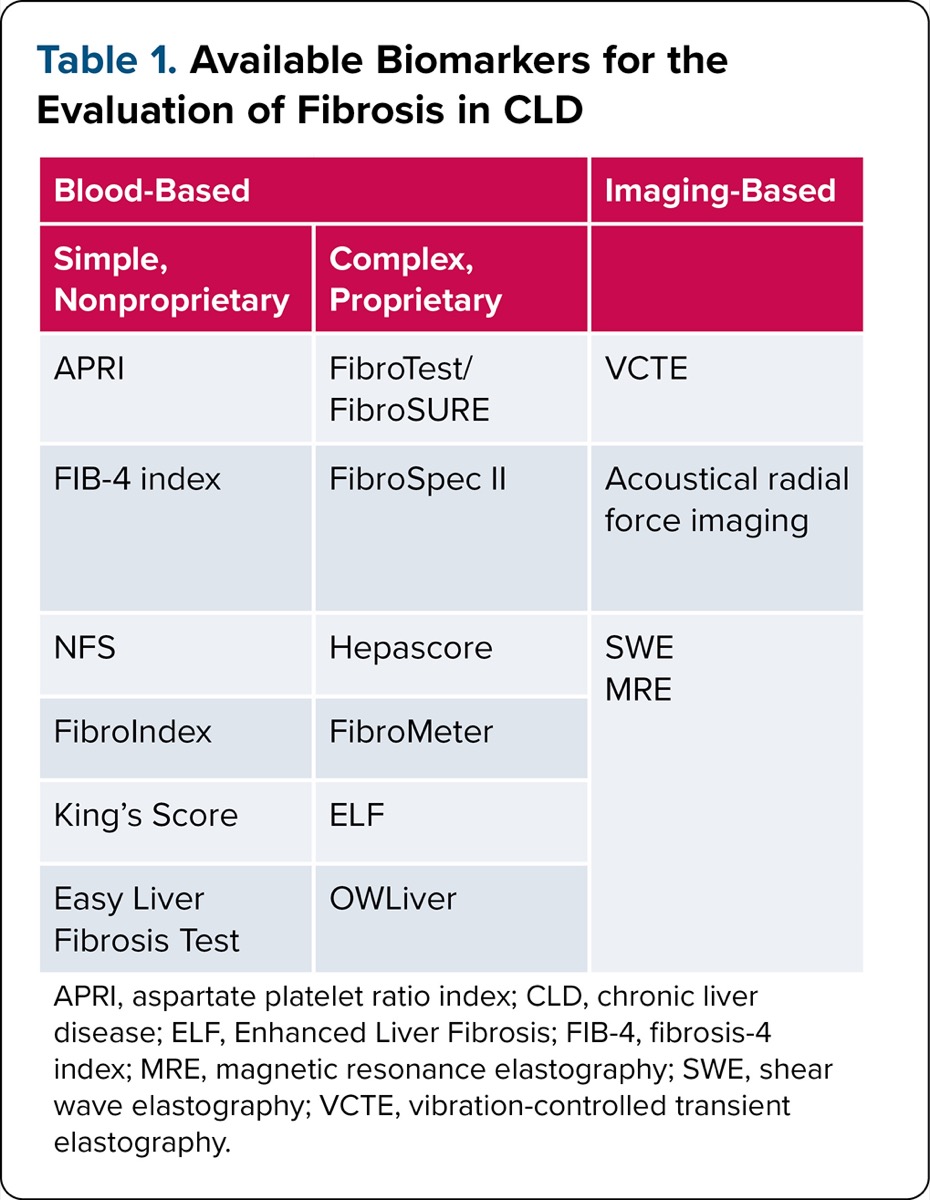
The American Association for the Study of Liver Diseases (AASLD) recently developed guidelines based on systematic reviews on the use of NILDAs to detect significant fibrosis, AF, and cirrhosis.2,6-9 NILDAs can be classified as either blood-based or imaging-based, and there is a wide array of available biomarkers. Blood-based NILDAs include routine tests, algorithms, and proprietary and patented tests and algorithms. They can be further broken down into simple, nonproprietary tests and more complex, proprietary tests (Table 1). Imaging-based NILDAs, based on elastography principles to assess liver stiffness, can be further stratified by their modality (Table 1).
| Table 1. Available Biomarkers for the Evaluation of Fibrosis in CLD | ||
| Blood-Based | Imaging-Based | |
|---|---|---|
| Simple, Nonproprietary | Complex, Proprietary | |
| APRI | FibroTest/FibroSURE | VCTE |
| FIB-4 index | FibroSpec II | Acoustical radial force imaging |
| NFS | Hepascore | SWE MRE |
| FibroIndex | FibroMeter | |
| King’s Score | ELF | |
| Easy Liver Fibrosis Test | OWLiver | |
| APRI, aspartate platelet ratio index; CLD, chronic liver disease; ELF, Enhanced Liver Fibrosis; FIB-4, fibrosis-4 index; MRE, magnetic resonance elastography; SWE, shear wave elastography; VCTE, vibration-controlled transient elastography. | ||
Interpreting both blood- and imaging-based NILDAs requires understanding several confounding factors. Specifically, blood-based NILDAs can be affected by age, splenectomy, gastrectomy, thrombocytopenia not related to portal hypertension, alcohol use, abnormal aspartate aminotransferase (AST) or alanine transaminase (ALT) values due to hepatic ischemia or acute liver injury, chronic kidney disease, malnutrition, inflammatory disorders, hemolysis, cholestatic diseases, postprandial hyperglycemia, extrahepatic fibrosing conditions, and autosickle cell crisis.2,10-19 Liver stiffness measurement (LSM) also can be affected by deposition diseases, such as amyloidosis, hepatic congestion in the setting of congestive heart failure or valvopathies, and hepatic infiltration in conditions such as leukemia.6
In the AASLD guidelines on NILDAs, the panel reviewed 9,447 articles published through April 2022. Among these studies, 286 addressed blood-based NILDAs2 and 240 addressed imaging-based NILDAs.6 Using the patient, intervention, comparison, and outcome method, the authors generated 21 guidance statements.2,6
Diagnostic performance is an important consideration in the use of NILDAs, and the application of standard statistical principles including sensitivity and specificity, both of which are independent of disease prevalence, is essential.
Noninvasive tests historically have had a much higher sensitivity than specificity, marking their utility in ruling out a condition. For many NILDAs, a low threshold has a higher sensitivity, and a higher threshold has a higher specificity. Conversely, both positive predictive value (PPV) and negative predictive value (NPV) are affected by disease prevalence, which is relevant, given factors such as patient selection in clinical practice because AF is less common in general practice than in hepatology practice, with estimates greater than 30% in the latter setting.2
To assess the accuracy of NILDAs across populations with varying prevalence, clinicians should use the diagnostic odds ratio. This ratio summarizes the odds of fibrosis in those with a positive test relative to that in those with a negative test2 and is a reliable estimate of a test’s accuracy, independent of the prevalence of the condition being tested.
Overall, careful interpretation of NILDAs is required to apply results correctly in clinical practice. A useful NILDA should have both a low threshold to rule out AF and a high threshold to rule in AF. Furthermore, when analyzing a NILDA, any result should be followed by assessing the test’s PPV and NPV in the population of interest before interpreting it or using the diagnostic odds ratio to assess its accuracy.
Both blood- and imaging-based NILDAs have limitations in their application, and the application of these tests is not uniform. The assessments were developed primarily to identify either AF or cirrhosis, not to assess significant fibrosis (=F2). Furthermore, many of these tests have up to 50% indeterminate results and are most accurate at the ends of the fibrosis spectrum, either F0 or F4. In addition, both blood- and imaging-based NILDAs were not developed to differentiate between adjacent fibrosis strata (eg, F2 from F3).2,6 Also, there are limited data and a lack of validation for using these tests as a measure to show improvement of fibrosis after disease-specific treatment. Further comparative and collaborative studies in these areas are needed.
Blood-Based NILDAs
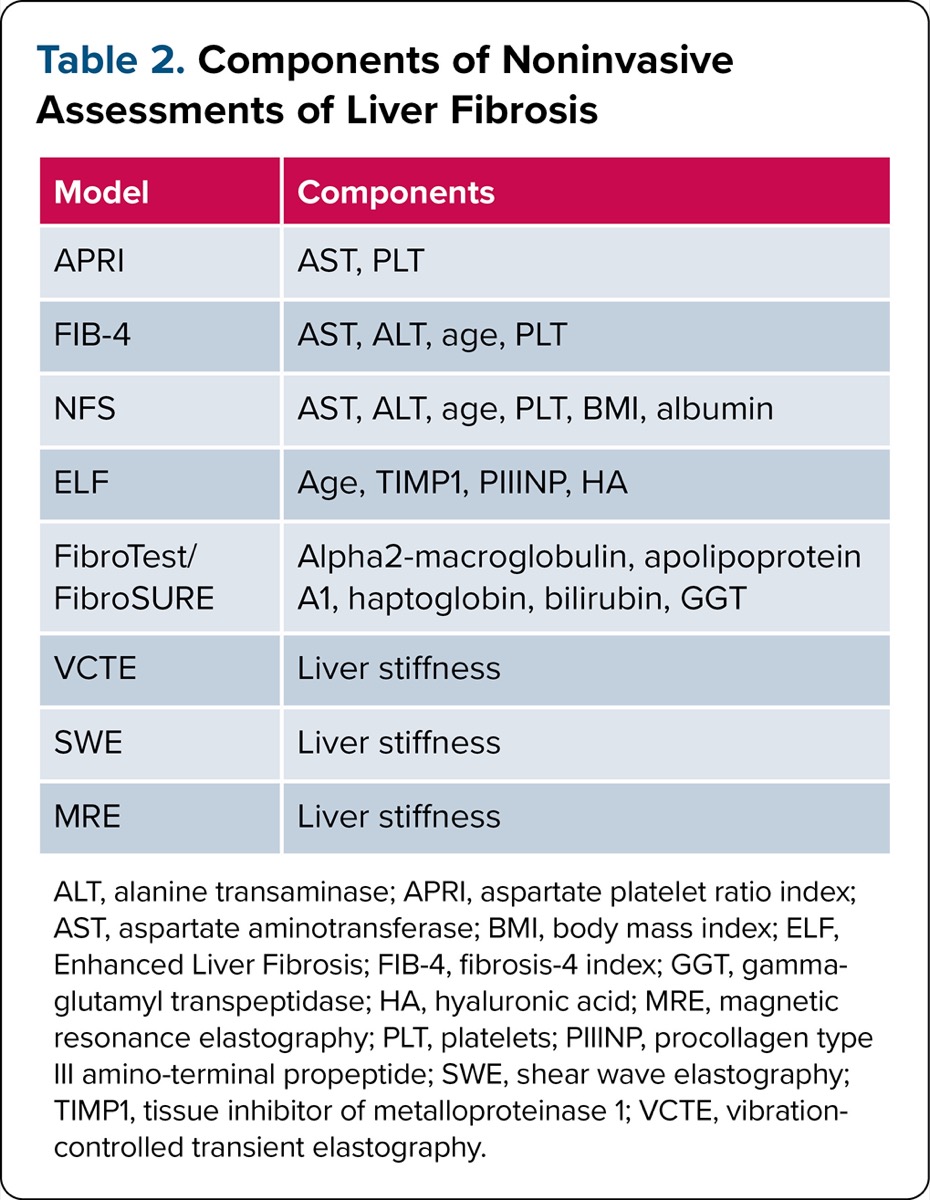
Numerous blood-based noninvasive tests have emerged over the past 2 decades (Table 1). Perhaps the 2 most studied are the Aspartate Platelet Ratio Index (APRI) and Fibrosis-4 Index (FIB-4).20-21 These NILDAs incorporate simple laboratory and demographic parameters; APRI is composed of AST and platelet count, whereas the FIB-4 also adds ALT and age (Table 2). Subsequent simple, noninvasive tests have been developed that often contain these core parameters and add additional metabolic parameters. These tests use readily accessible inputs and are easily computed with online calculators. However, simple scores can be misleading in patients at extremes of age or who have confounding explanations for abnormal laboratory inputs, such as acute hepatitis or an alternative cause of thrombocytopenia. More complex, proprietary tests have been developed that often include direct molecular markers of fibrosis but are more costly and less accessible.
| Table 2. Components of Noninvasive Assessments of Liver Fibrosis | |
| Model | Components |
|---|---|
| APRI | AST, PLT |
| FIB-4 | AST, ALT, age, PLT |
| NFS | AST, ALT, age, PLT, BMI, albumin |
| ELF | Age, TIMP1, PIIINP, HA |
| FibroTest/FibroSURE | Alpha2-macroglobulin, apolipoprotein A1, haptoglobin, bilirubin, GGT |
| VCTE | Liver stiffness |
| SWE | Liver stiffness |
| MRE | Liver stiffness |
| ALT, alanine transaminase; APRI, aspartate platelet ratio index; AST, aspartate aminotransferase; BMI, body mass index; ELF, Enhanced Liver Fibrosis; FIB-4, fibrosis-4 index; GGT, gamma-glutamyl transpeptidase; HA, hyaluronic acid; MRE, magnetic resonance elastography; PLT, platelets; PIIINP, procollagen type III amino-terminal propeptide; SWE, shear wave elastography; TIMP1, tissue inhibitor of metalloproteinase 1; VCTE, vibration-controlled transient elastography. | |
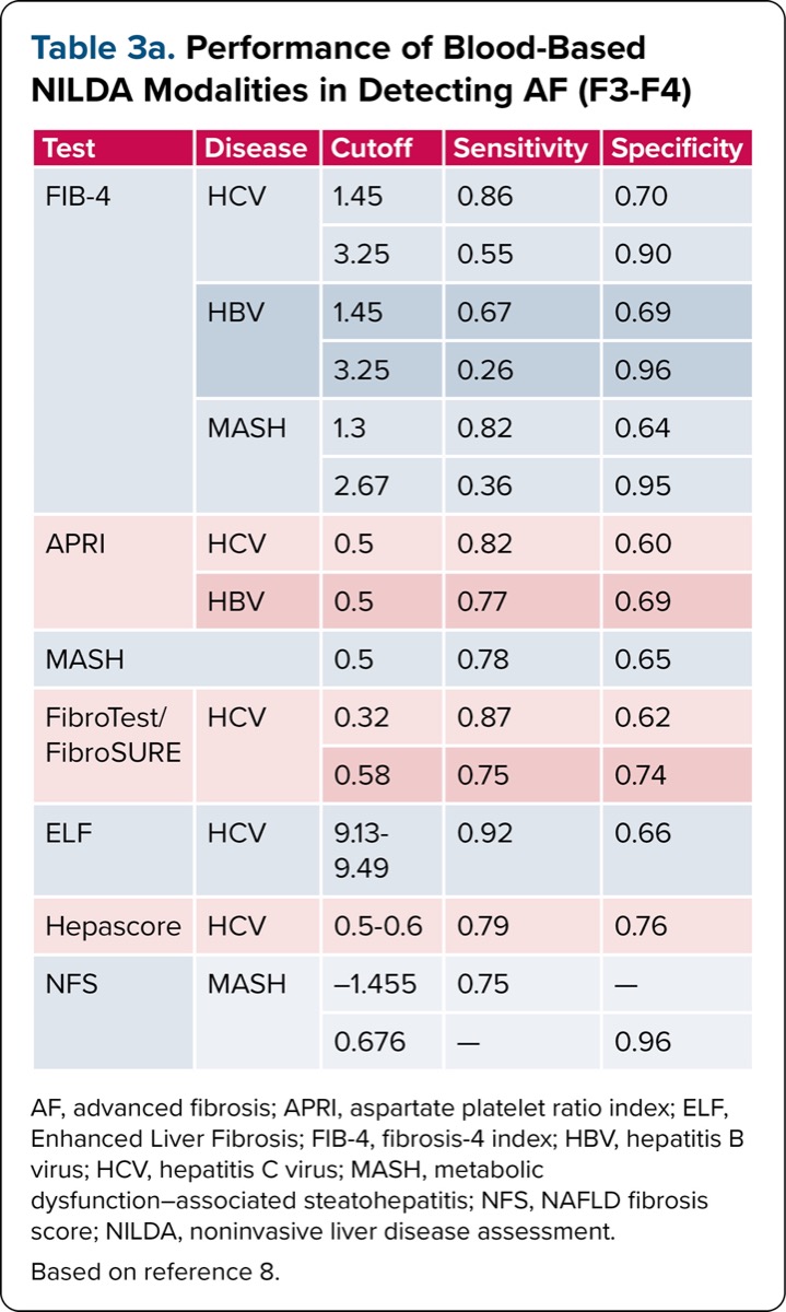
The 2025 AASLD guidelines on blood-based NILDAs recommend using simple blood-based noninvasive scores, such as APRI or FIB-4, over proprietary tests as an initial test to identify fibrosis stage in patients with chronic viral hepatitis.2 In the AASLD guideline and systematic review, FIB-4 cutoffs of 1.45/3.25 had a sensitivity/specificity of 0.86/0.70 and 0.55/0.90, respectively, for the detection of AF in HCV (Table 3a).2,8 In chronic HBV, the FIB-4 had decreased sensitivity (0.67) for the diagnosis of AF at the lower cutoff of 1.45 but maintained high specificity (0.96) at the upper cutoff of 3.25.
| Table 3a. Performance of Blood-Based NILDA Modalities in Detecting AF (F3-F4) | ||||
| Test | Disease | Cutoff | Sensitivity | Specificity |
|---|---|---|---|---|
| FIB-4 | HCV | 1.45 | 0.86 | 0.70 |
| 3.25 | 0.55 | 0.90 | ||
| HBV | 1.45 | 0.67 | 0.69 | |
| 3.25 | 0.26 | 0.96 | ||
| MASH | 1.3 | 0.82 | 0.64 | |
| 2.67 | 0.36 | 0.95 | ||
| APRI | HCV | 0.5 | 0.82 | 0.60 |
| HBV | 0.5 | 0.77 | 0.69 | |
| MASH | 0.5 | 0.78 | 0.65 | |
| FibroTest/ FibroSURE | HCV | 0.32 | 0.87 | 0.62 |
| 0.58 | 0.75 | 0.74 | ||
| ELF | HCV | 9.13-9.49 | 0.92 | 0.66 |
| Hepascore | HCV | 0.5-0.6 | 0.79 | 0.76 |
| NFS | MASH | –1.455 | 0.75 | — |
| 0.676 | — | 0.96 | ||
| AF, advanced fibrosis; APRI, aspartate platelet ratio index; ELF, Enhanced Liver Fibrosis; FIB-4, fibrosis-4 index; HBV, hepatitis B virus; HCV, hepatitis C virus; MASH, metabolic dysfunction–associated steatohepatitis; NFS, NAFLD fibrosis score; NILDA, noninvasive liver disease assessment. Based on reference 8. | ||||
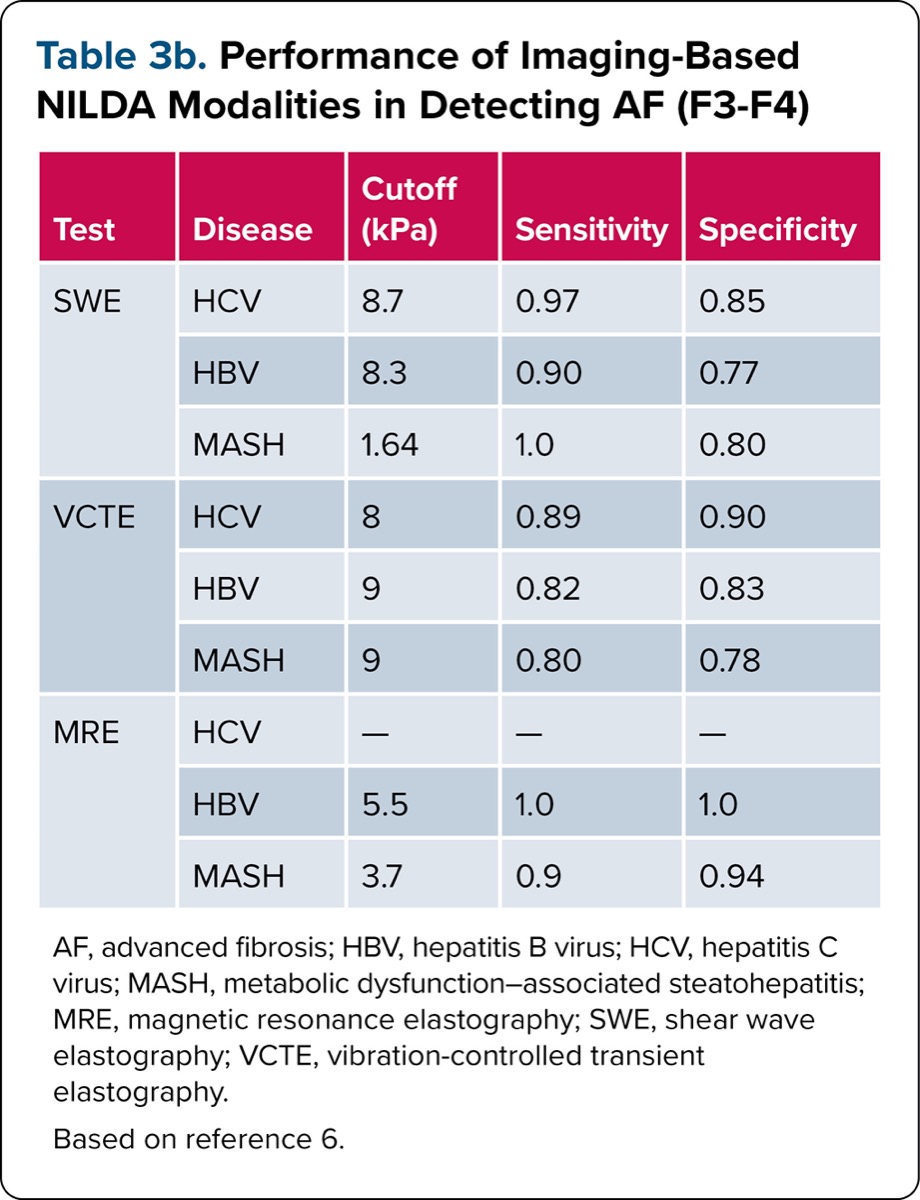
| Table 3b. Performance of Imaging-Based NILDA Modalities in Detecting AF (F3-F4) | ||||
| Test | Disease | Cutoff (kPa) | Sensitivity | Specificity |
|---|---|---|---|---|
| SWE | HCV | 8.7 | 0.97 | 0.85 |
| HBV | 8.3 | 0.90 | 0.77 | |
| MASH | 1.64 | 1.0 | 0.80 | |
| VCTE | HCV | 8 | 0.89 | 0.90 |
| HBV | 9 | 0.82 | 0.83 | |
| MASH | 9 | 0.80 | 0.78 | |
| MRE | HCV | — | — | — |
| HBV | 5.5 | 1.0 | 1.0 | |
| MASH | 3.7 | 0.9 | 0.94 | |
| AF, advanced fibrosis; HBV, hepatitis B virus; HCV, hepatitis C virus; MASH, metabolic dysfunction–associated -steatohepatitis; MRE, magnetic resonance elastography; SWE, shear wave elastography; VCTE, vibration-controlled transient elastography. Based on reference 6. | ||||
All noninvasive tests should be used prior to initiating therapy in patients with chronic HCV or chronic HBV because there are no validated NILDA cutoffs that correlate with fibrosis stage after sustained virologic response in HCV or viral suppression in HBV. Treatments of the underlying liver disease improve AST and ALT values, resulting in underestimation of fibrosis stage by blood-based NILDAs.
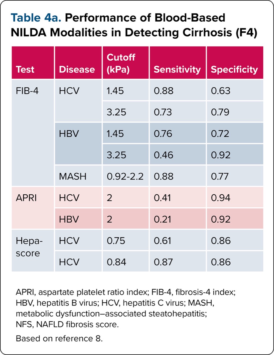
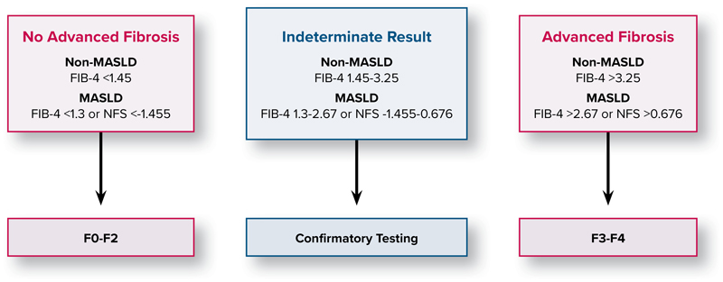
In patients with MASLD, the guidelines also recommend using simple blood-based noninvasive tests over complex, proprietary tests to detect AF (F3-F4). A systematic review of 32 studies evaluating the FIB-4 found a pooled specificity of 0.94 for upper cutoffs (FIB-4=2.67 and =3.25 in diagnosing AF and cirrhosis) (Tables 3a and 4a).2 The NAFLD fibrosis score (NFS) is a simple blood-based NILDA that incorporates body mass index (BMI), diabetes status, and albumin in addition to the FIB-4 parameters.22 In the guideline panel’s review of available studies evaluating NFS, a cutoff of less than –1.455 yielded a sensitivity of 0.75 (95% CI, 0.61-0.81) for ruling out AF, while a cutoff of greater than 0.676 had a specificity of 0.96 (95% CI, 0.93-0.98) for diagnosis of AF. However, 33.5% of patients fell within the indeterminate range.
| Table 4a. Performance of Blood-Based NILDA Modalities in Detecting Cirrhosis (F4) | ||||
| Test | Disease | Cutoff (kPa) | Sensitivity | Specificity |
|---|---|---|---|---|
| FIB-4 | HCV | 1.45 | 0.88 | 0.63 |
| 3.25 | 0.73 | 0.79 | ||
| HBV | 1.45 | 0.76 | 0.72 | |
| 3.25 | 0.46 | 0.92 | ||
| MASH | 0.92-2.2 | 0.88 | 0.77 | |
| APRI | HCV | 2 | 0.41 | 0.94 |
| HBV | 2 | 0.21 | 0.92 | |
| Hepa-score | HCV | 0.75 | 0.61 | 0.86 |
| HCV | 0.84 | 0.87 | 0.86 | |
| APRI, aspartate platelet ratio index; FIB-4, fibrosis-4 index; HBV, hepatitis B virus; HCV, hepatitis C virus; MASH, metabolic dysfunction–associated steatohepatitis; NFS, NAFLD fibrosis score. Based on reference 8. | ||||
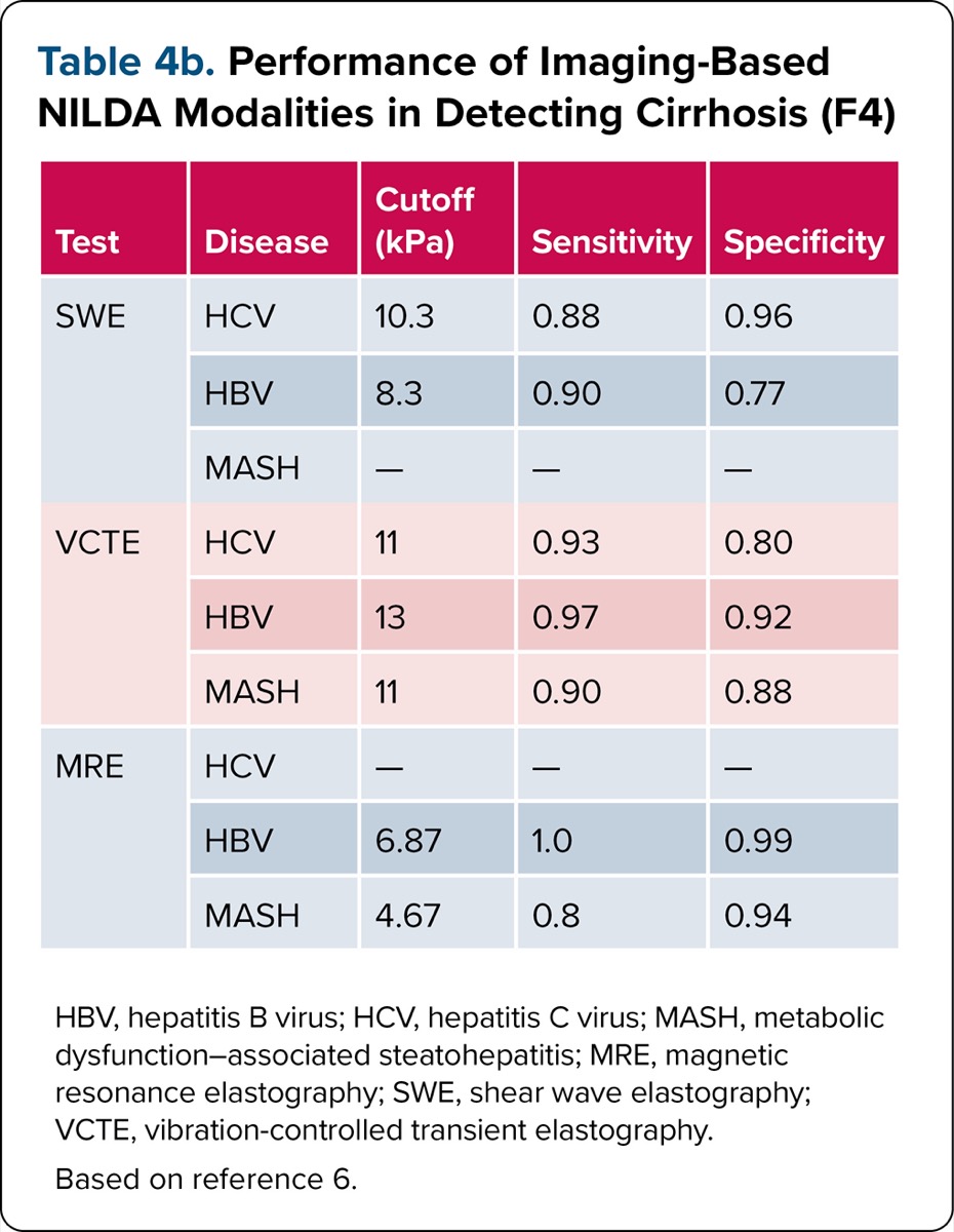
| Table 4b. Performance of Imaging-Based NILDA Modalities in Detecting Cirrhosis (F4) | ||||
| Test | Disease | Cutoff (kPa) | Sensitivity | Specificity |
|---|---|---|---|---|
| SWE | HCV | 10.3 | 0.88 | 0.96 |
| HBV | 8.3 | 0.90 | 0.77 | |
| MASH | — | — | — | |
| VCTE | HCV | 11 | 0.93 | 0.80 |
| HBV | 13 | 0.97 | 0.92 | |
| MASH | 11 | 0.90 | 0.88 | |
| MRE | HCV | — | — | — |
| HBV | 6.87 | 1.0 | 0.99 | |
| MASH | 4.67 | 0.8 | 0.94 | |
| HBV, hepatitis B virus; HCV, hepatitis C virus; MASH, metabolic dysfunction–associated steatohepatitis; MRE, magnetic resonance elastography; SWE, shear wave elastography; VCTE, vibration-controlled transient elastography. Based on reference 6. | ||||
Figure 1 and Table 3a highlight the FIB-4 and NFS thresholds proposed by the AASLD guideline panel for fibrosis staging.2 To identify AF in MASLD, the guideline panel suggests that a sequential combination of blood-based NILDAs may be considered rather than a single test, although the quality of evidence is low. They further note that blood-based biomarkers should not be used to assess steatosis in MASLD patients.
Most NILDA studies have been performed in patients with viral hepatitis or MASLD. There was insufficient evidence for the guideline panel to recommend blood-based NILDAs for fibrosis assessment in patients with alcohol-associated liver disease or cholestatic liver disease. In pediatric patients with CLD, the guideline panel suggests that simple blood-based NILDAs, such as APRI or FIB-4, should be used for the diagnosis of AF. They note that blood-based NILDAs are not suitable to discriminate earlier stages of fibrosis and that and blood-based NILDAs incorporating age as a parameter, including FIB-4, do not perform as well in children as adults.
Given the frequency of indeterminate results with blood-based NILDAs, the panel suggests that a combination of noninvasive tests performed sequentially may perform better than a single test for diagnosing significant fibrosis or cirrhosis. Blood-based NILDAs have high sensitivity for ruling out AF but can have limited PPV for confident diagnoses. In addition, blood-based noninvasive models frequently categorize patients into indeterminate ranges, which necessitates additional testing to assess fibrosis stage. NILDAs have difficulty differentiating between adjacent stages of disease, which limits their use in tracking longitudinal changes in fibrosis. Thus, the AASLD advises against using blood-based noninvasive tests to follow progression, stability, or regression in histologic stage in patients with CLD.
Imaging-Based NILDAs
Broadly speaking, there are 3 imaging-based NILDA techniques available for widespread use (Table 1). These include vibration-controlled transient elastography (VCTE), shear wave elastography (SWE), and magnetic resonance elastography (MRE). A key advantage of imaging-based NILDAs over blood-based NILDAs is that in addition to detecting fibrosis, these modalities also enable the quantification of hepatic steatosis. Among the 3 techniques, VCTE is the best validated, most widely available, and most used modality in clinical practice, followed by SWE and MRE.
VCTE and SWE are ultrasound-based techniques that detect liver stiffness by measuring the speed with which shear waves transit through the parenchyma.23 The key difference in technique between the 2 is the method used to generate shear waves. VCTE uses vibration to generate shear waves, whereas in SWE, the absorption of an acoustic pulse by tissue results in generation of shear waves.24 MRE uses mechanical shear waves generated by a plastic disc placed over the patient’s upper-right quadrant and connected to an external wave generator, referred to as an active driver.25 The speed at which this generates shear waves across the liver is then detected using MRI techniques, allowing for the evaluation of cross-sectional imaging as well as an assessment of liver stiffness.
The 2025 AASLD guidelines recommend use of imaging-based NILDAs across the entire spectrum of chronic liver diseases to detect significant fibrosis (F2-F4), AF (F3-F4), and cirrhosis (F4). Imaging-based NILDAs have excellent sensitivity and specificity to assess for AF (Table 3b) and cirrhosis (Table 4b).6 The guidelines also recommend the use of imaging-based NILDA to detect AF and cirrhosis in patients with alcohol-associated liver disease and chronic cholestatic liver diseases.6 Of note, much of the data on viral hepatitis were collected from viremic patients and, as such, the appropriate use of NILDAs for patients who have completed antiviral therapy is poorly defined.6 Overall, evidence for the use of imaging-based NILDAs is not as robust in these disease states.
The guidelines further specify that either ultrasound-based elastography (VCTE or SWE) or MRE can be used for noninvasive detection of fibrosis. Choice of imaging modality should be based on local availability, expertise, need for cross-sectional imaging, and patient factors that may confound the accuracy of ultrasound-based testing and, thus, should be considered in this diagnostic decision.6 Patient factors such as ascites and obesity have the greatest effect on LSM accuracy in VCTE, followed by SWE,6,26 which may support use of MRE in these contexts. The XL VCTE probe can counter VCTE-LSM failure in many cases of obesity, but in patients with extreme obesity (BMI =40 kg/m2), the XL probe may still produce inaccurate results.
The totality of the available data shows that imaging-based NILDAs generally outperform blood-based diagnostics.6 On these grounds, the AASLD panel recommends that, if available, imaging-based techniques be incorporated into the initial fibrosis staging of adults with CLD. Although imaging-based techniques are considered superior, the guidelines support either concomitant or sequential combination of blood- and imaging-based techniques to improve the accuracy of noninvasive fibrosis detection.6 This strategy was implemented into the 2023 AASLD practice guidance on the clinical assessment and management of MASLD, which recommends the use of FIB-4 as a primary risk assessment, followed by VCTE.27 The easy accessibility of most blood-based NILDAs allows for convenient implementation as an initial screening tool across a variety of clinical settings.
The AASLD does not recommend stand-alone imaging-based NILDAs for longitudinal monitoring of regression or progression of fibrosis. It also is important to understand the effect that patient factors—such as obesity, alcohol use, viremia, and more—have on LSM when interpreting these results. For example, LSM is less reliable when ALT or AST are greater than 100 U/mL, which can be important when evaluating patients in acute or acute-on-chronic disease states, such as in the setting of acute alcohol– associated hepatitis.28 Although there are promising data on the use of imaging-based NILDAs to track changes in fibrosis longitudinally, the evidence remains insufficient to broadly support the practice.6
In the detection of steatosis, blood-based NILDAs do not perform as well as imaging-based NILDAs. Transient elastography–controlled attenuation parameter (TE-CAP) has good diagnostic accuracy to grade steatosis and typically is used first-line in screening for steatosis given its wide availability and convenience as a point-of-care test.6 MRI proton density fat fraction has superior accuracy to TE-CAP, but it is more expensive and less widely available than its ultrasound-based counterparts.6
Within the pediatrics population, there is insufficient evidence to recommend one imaging-based NILDA over another. There are still limited data evaluating imaging-based NILDAs in the pediatric population, but in practice, clinicians use both MRE and VCTE to quantify steatosis in children with both metabolic dysfunction–associated steatohepatitis and cystic fibrosis.
Conclusion
With the advent of NILDAs, clinicians now have less expensive and safer alternatives to liver biopsy for the evaluation of CLD.6 The 2025 AASLD guidelines on blood-based and imaging-based NILDAs provide a formal roadmap for their implementation based on 21 guidance statements. As depicted in Figure 2, clinicians can use a sequential approach to accurately assess the degree of fibrosis and reduce the pool of patients who, ultimately, will require liver biopsy.
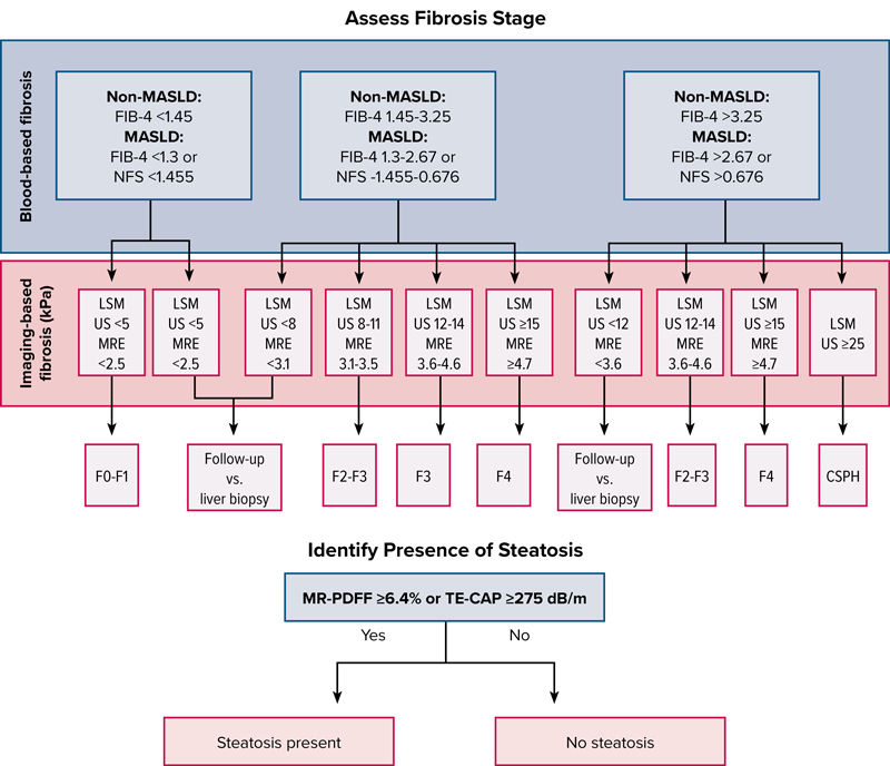
FIB-4 and NFS have good specificity for AF and serve as initial “rule-out” tests. Since these are calculated using commonly accessible parameters, they are universally accessible across all clinical settings. Imaging-based NILDAs can then be implemented to further stratify patients by their degree of fibrosis. The use of ultrasound- and MRI-based techniques also allows for the quantification of steatosis.
Despite their utility, NILDAs are imperfect. There remains a subset of patients who require liver biopsy for diagnosis due to indeterminant results, although this subset of patients is much smaller now that NILDA use is more widespread. There are also limited data to correlate these assessments with post-treatment disease states (eg, HCV after direct-acting antivirals, HBV with suppression, and autoimmune hepatitis after therapy). Access to imaging-based NILDAs, both in local availability of equipment as well as the expertise to operate it, remains limited. Ultrasound-based modalities, specifically, can be quite operator- dependent, despite being the most used imaging-based NILDA.
The patient population most coveted for clinical trials remains those with F2-F3 because these patients can be treated most effectively with pharmacotherapeutics. NILDAs, like most biomarkers, are designed to detect the extremes of disease and, as such, play a limited role in capturing moderate fibrosis.
A tremendous amount of progress has been made in the field to develop effective modes of noninvasive evaluation of liver disease. There are newer models moving beyond the NILDAs covered in the AASLD guidelines that may address these limitations.29 The future continues to hold a great deal of promise, from artificial intelligence and machine learning technologies that may further improve diagnostic techniques, to advances in hardware and software efficiencies to facilitate easier access and implementation of existing NILDAs.6
References
- Hagström H, Nasr P, Ekstedt M, et al. Fibrosis stage but not NASH predicts mortality and time to development of severe liver disease in biopsy-proven NAFLD. J Hepatol. 2017;67(6):1265-1273.
- Sterling RK, Patel K, Duarte-Rojo A, et al. AASLD Practice Guideline on blood-based non-invasive liver disease assessments of hepatic fibrosis and steatosis. Hepatology. 2025;81(1):321-357.
- Kleiner DE, Brunt EM, Van Natta M, et al. Design and validation of a histological scoring system for nonalcoholic fatty liver disease. Hepatology. 2005;41(6):1313-1321.
- Brunt EM, Janney CG, Di Bisceglie AM, et al. Nonalcoholic steatohepatitis: a proposal for grading and staging the histological lesions. Am J Gastroenterol. 1999;94(9):2467-2474.
- Rockey DC, Caldwell SH, Goodman ZD, et al. Liver biopsy. Hepatology. 2008;49(3):1017-1044.
- Sterling RK, Duarte-Rojo A, Patel K, et al. AASLD Practice Guideline on imaging-based non-invasive liver disease assessments of hepatic fibrosis and steatosis. Hepatology. 2025;81(2):672-724.
- Sterling RK, Asrani SK, Levine D, et al. AASLD Practice Guideline on non-invasive liver disease assessments of portal hypertension. Hepatology. 2025;81(3):1060-1085.
- Patel K, Asrani SK, Fiel MI, et al. Accuracy of blood-based biomarkers for staging liver fibrosis in chronic liver disease: a systematic review supporting the AASLD Practice Guideline. Hepatology. 2025;81(1):358-379.
- Duarte-Rojo A, Taouli B, Leung DH, et al. Imaging-based noninvasive liver disease assessment for staging liver fibrosis in chronic liver disease: a systematic review supporting the AASLD Practice Guideline. Hepatology. 2025 Feb 1;81(2):725-748.
- Nguyen-Khac E, Thiele M, Voican C, et al. Non-invasive diagnosis of liver fibrosis in patients with alcohol-related liver disease by transient elastography: an individual patient data meta- analysis. Lancet Gastroenterol Hepatol. 2018;3(9):614-625.
- Petta S, Wong VWS, Bugianesi E, et al. Impact of obesity and alanine aminotransferase levels on the diagnostic accuracy for advanced liver fibrosis of noninvasive tools in patients with nonalcoholic fatty liver disease. Am J Gastroenterol. 2019;114(6):916-928.
- Wong GL, Wong VW, Choi PC, et al. Increased liver stiffness measurement by transient elastography in severe acute exacerbation of chronic hepatitis B. J Gastroenterol Hepatol. 2009;24(6):1002-1007.
- Liu CH, Liang CC, Huang KW, et al. Transient elastography to assess hepatic fibrosis in hemodialysis chronic hepatitis C patients. Clin J Am Soc Nephrol. 2011;6(5):1057-1065.
- Taneja S, Borkakoty A, Rathi S, et al. Assessment of liver fibrosis by transient elastography should be done after hemodialysis in end stage renal disease patients with liver disease. Dig Dis Sci. 2017;62(11):3186-3192.
- Schmoyer CJ, Kumar D, Gupta G, et al. Diagnostic accuracy of noninvasive tests to detect advanced hepatic fibrosis in patients with hepatitis C and end-stage renal disease. Clin Gastroenterol Hepatol. 2020;18(10):2332-2339.e1.
- Vuppalanchi R, Weber R, Russell S, et al. Is fasting necessary for individuals with nonalcoholic fatty liver disease to undergo vibration-controlled transient elastography? Am J Gastroenterol. 2019;114(6):995-997.
- Murawaki Y, Idobe Y, Ikuta Y, et al. Influence of a history of gastrectomy for gastric cancer on serum hyaluronan concentration in normal individuals and patients with chronic liver disease. Hepatol Res. 1998;10(3):248-254.
- Su Y, Gu H, Weng D, et al. Association of serum levels of laminin, type IV collagen, procollagen III N-terminal peptide, and hyaluronic acid with the progression of interstitial lung disease. Medicine. 2017;96(18):e6617-e6617.
- Koh C, Turner T, Zhao X, et al. Liver stiffness increases acutely during sickle cell vaso-occlusive crisis. Am J Hematol. 2013;88(11):E250-E254.
- Wai CT, Greenson JL, Fontana RJ, et al. A simple non-invasive index can predict both significant fibrosis and cirrhosis in patients with chronic hepatitis C. Hepatology 2003;38(2):518-526.
- Sterling RK, Lissen E, Clumeck N, et al. Development of a simple noninvasive index to predict significant fibrosis in patients with HIV/HCV coinfection. Hepatology. 2006;43(6):1317-1325.
- Angulo P, Hui JM, Marchesini G, et al. The NAFLD fibrosis score: a noninvasive system that identifies liver fibrosis in patients with NAFLD. Hepatology. 2007;45(4):846-854.
- Wong VWS, Vergniol J, Wong GLH, et al. Diagnosis of fibrosis and cirrhosis using liver stiffness measurement in nonalcoholic fatty liver disease. Hepatology. 2010;51(2):454-462.
- Kennedy P, Wagner M, Castéra L, et al. Quantitative elastography methods in liver disease: current evidence and future directions. Radiology. 2018;286(3):738-763.
- Akkaya HE, Erden A, Kuru öz D, et al. Magnetic resonance elastography: basic principles, technique, and clinical applications in the liver. Diagn Interv Radiol. 2018;24(6):328-335.
- Sigrist RMS, Liau J, Kaffas AE, et al. Ultrasound elastography: review of techniques and clinical applications. Theranostics. 2017;7(5):1303-1329.
- Rinella ME, Neuschwander-Tetri BA, Siddiqui MS, et al. AASLD Practice Guidance on the clinical assessment and management of nonalcoholic fatty liver disease. Hepatology. 2023;77(5):1797-1835.
- Altamirano J, Qi Q, Choudhry S, et al. Non-invasive diagnosis: non-alcoholic fatty liver disease and alcoholic liver disease. Transl Gastroenterol Hepatol. 2020;5:31.
- Grady JT, Cyrus JW, Sterling RK. Novel noninvasive tests for liver fibrosis: moving beyond simple tests in metabolic dysfunction-associated steatotic liver disease. Clin Gastroenterol Hepatol. Published online May 30, 2025. doi:10.1016/j.cgh.2025.02.035
Copyright © 2025 McMahon Publishing, 545 West 45th Street, New York, NY 10036. Printed in the USA. All rights reserved, including the right of reproduction, in whole or in part, in any form.
Download to read this article in PDF document:![]() In Lieu of Biopsy: A Review of Noninvasive Liver Disease Assessments
In Lieu of Biopsy: A Review of Noninvasive Liver Disease Assessments


