The tripod grasper was originally developed to retrieve colon polyps. Given its versatile design, it soon became evident that this device also was useful for removing other types of objects.
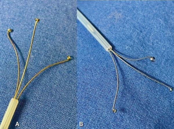
The tripod grasper consists of a 3-pronged wire inside a catheter. Once the prongs are extended out of the cannula, they open like a bird claw (Figure 1A-B). Of note, in contrast to real claws, the tips are rounded to avoid damaging the mucosa or grasping unwanted tissue. The objective is to get the object inside the claws and remove it without pulling off any adjacent tissue.
We describe 2 cases in which the tripod grasper facilitated endoscopic removal of a foreign object from patients with a history of esophageal conditions.
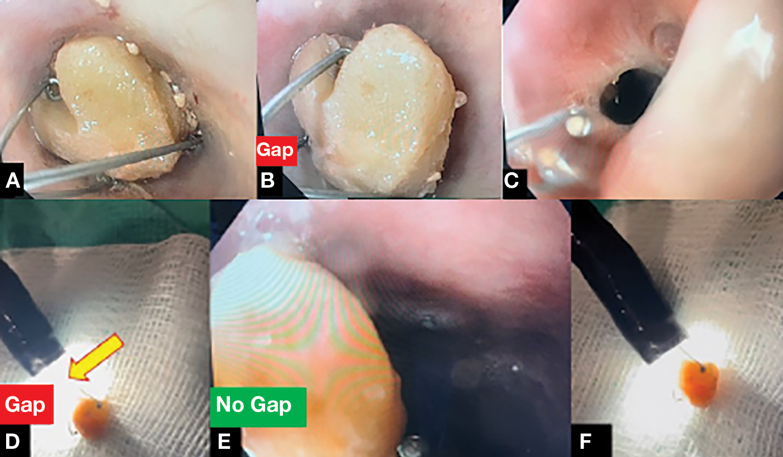
In case 1, an older patient with a history of previous lye ingestion and complex esophageal strictures presented with food impaction lasting 24 hours (Figure 2A-F). The patient had known esophageal strictures since childhood and was on a regular dilation program. Use of a cap was not possible for this patient because the esophagus was stenosed due to previous lye injury. Indeed, use of caps is difficult or impossible in any patient with a fibrotic esophagus due to radiation esophagitis, old lye injury, or tight anastomosis because of the narrowed lumen.
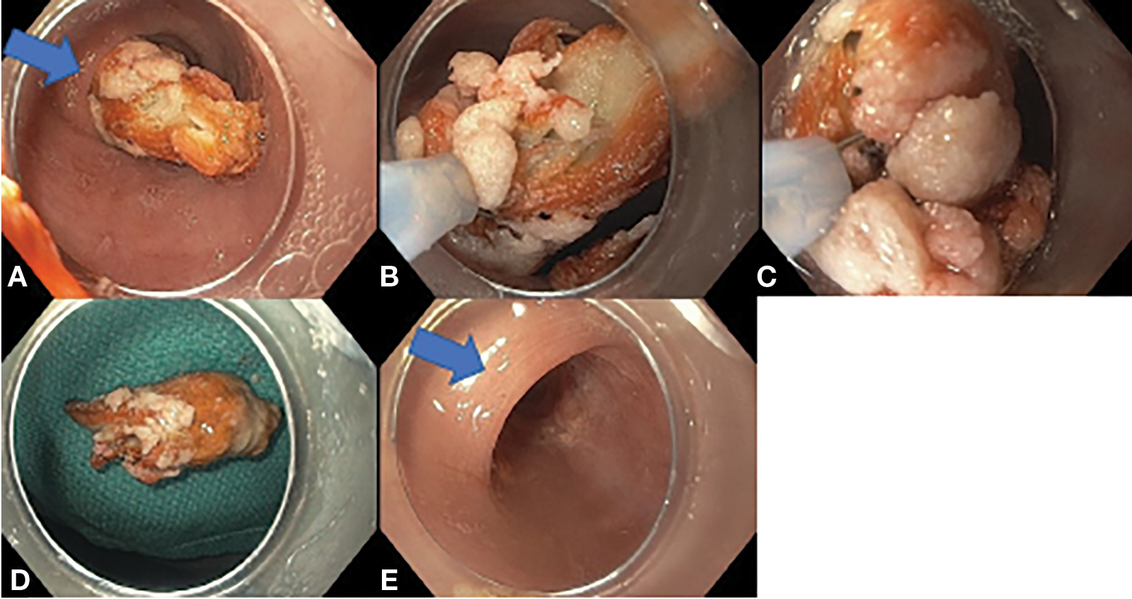
Case 2 was a young patient with a history of long-standing eosinophilic esophagitis that mainly had been treated with proton pump inhibitors, who presented with an impaction of chicken meat (Figure 3A-E).
Herein we present some tips and tricks on using the tripod grasper.
First, inspect the motility of the esophagus and the interplay of the foreign body with the stenosis and the stricture. Is the esophagus elastic or stiff? Is the foreign body impacted or does it move up and down? It is helpful to use some water to further determine the interplay of the foreign body and the stricture.
Second, evaluate the mucosa. We want to prevent further damage during manipulation during the extraction maneuvers. Is the mucosa macerated due to long-standing pressure damage from the foreign body? Has the patient developed a laceration due to nausea, vomiting, or retching?
Third, proceed to remove the foreign body. It is important not to fully open or expose the entire tripod grasper as you approach (Figure 2A) because it may get entangled against the walls of the stenotic esophagus. However, once you are within reach of the foreign body, partially open the grasper. With the partially opened grasper, advance the scope toward the foreign body and slowly continue to fully expose (open) the grasper’s legs. When you are on top of the foreign body, start closing the grasper, while simultaneously pushing toward the foreign body (Figure 2B). Closing without pushing will result in an inefficient grasp, and the foreign body may fall off the grasper.
Fourth, once the foreign body is tightly grasped, proceed to pull the foreign body toward the tip of the scope, to avoid a gap between the scope tip and grasped foreign body (Figure 2E-F). This is called the pathfinder trick because the scope will serve as guide and “clear the way” for the foreign body on its tip.
If there is a distance between the scope and tripod grasper with the foreign body (Figure 2D, yellow arrow showing the gap), the foreign body may fall off at areas of the stenosis, such as the upper esophageal sphincter or additional esophageal stenosis or fibrotic rings (Figure 2C).
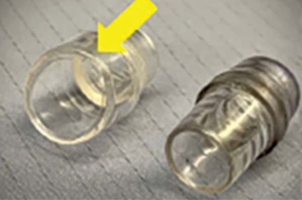
In the patient with chicken meat impaction, a first attempt was made to remove it with a distal transparent cap, but a fibrotic ring impeded the dislodgment of the meat (Figure 3A-B). Thus, a combination approach was used. The tripod grasper was used to catch the meat, while the cap fulfilled 2 functions, keeping the proximal part of the meat inside and being a “pathfinder.” Because the cap (Figure 4, yellow arrow) is wider than the endoscope, it “opens the way,” that is, “keeping the path free” for the caught object or meat, being especially useful when traversing stenosis, webs, or coming out of the upper esophageal sphincter.
One last trick: While removing the scope with the foreign body, try to gently twist the scope, in a corkscrew-type way, to adapt to esophageal tortuosity and, thus, avoid having the foreign body bounce off the wall or mucosal strictures.
Images courtesy of EndoCollab. See endocollab.com for more information, including videos and lectures on these and many other practical endoscopy tricks.
{RELATED-HORIZONTAL}


